
Our Research
“I think, therefore I am.” (Cogito, ergo sum)
We are engaged in designing and developing new types of materials that may find application in agriculture and the environment. For this, we use a “structure-and-function” approach to our materials research. Our current research is focused on increasing the nutrient use efficiency (NUE) of nitrogen (N) and phosphorus (P) in agricultural systems.
For this, we explore the large material space at the nano- to micro-scale, typically ranging from porous inorganic or organic materials to composites, hybrid and nano-biohybrid materials. In doing so, we get an en route opportunity to examine crystallisation in non-equilibrium conditions, (e⁻) transfers at mineral–water interfaces, light (hν)-induced processes at mineral–water–bio interfaces, and the adsorption and release of nutrients (mineral and organic) in a complex matrix (soil).
Apart from this, in collaboration with other groups, we conduct research to integrate artificial intelligence and machine learning (AI-ML) with soil diagnostic data and agronomic intelligence for on-farm decision-making in fertilizer use. In terms of research methods, we are primarily an experimental group, but we are also developing capabilities in computational chemistry and AI-ML to power our materials discovery and fertilizer research. In policy terms, our research falls largely within circular economy and sustainability work.
Theme A
Materials Design & Synthesis
Bio-mimetically inspired
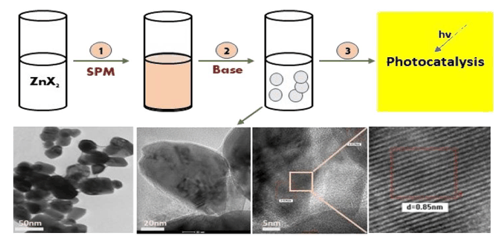
The current materials design and synthesis efforts are directed at finding sustainable ways to obtain materials with desired characteristics. In the same line of effort, and as a part of ligand field/design theory, we are attempting to extend the biomimetics approach to control the shape anisotropy of nanosized metal oxide (FeₓOᵧ, ZnO, CuO, etc.) crystals by replacing fine chemicals (amino acids, peptides, surfactants and polymers) as ligands with bio-based complex lignocellulosic substances from agro-industrial wastes.
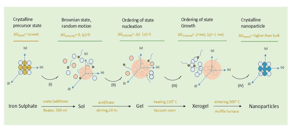
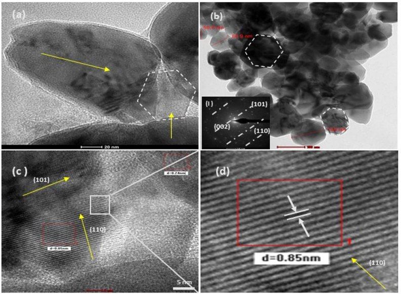
We found in our studies that the complex field of ligands and their structural properties under sol-gel conditions can induce shape anisotropy in nanocrystals that may remain preserved after heat treatment (calcination). These results have been compared with analytical-grade surfactants and ligands such as CTAB and methyl cellulose. These findings raise a further mechanistic question: how can the complex environment of ligands produce results similar to those of pure analytical analogues?
Source: L. M. Verma et al., Nanoscale Adv. 6, 155–169 (2024); L. M. Verma et al., J. Mater. Chem. A 13, 33365–33384 (2025). doi:10.1039/D3NA00596H ↗
Theme B
Non-equilibrium Crystallisation
Ligand field theories
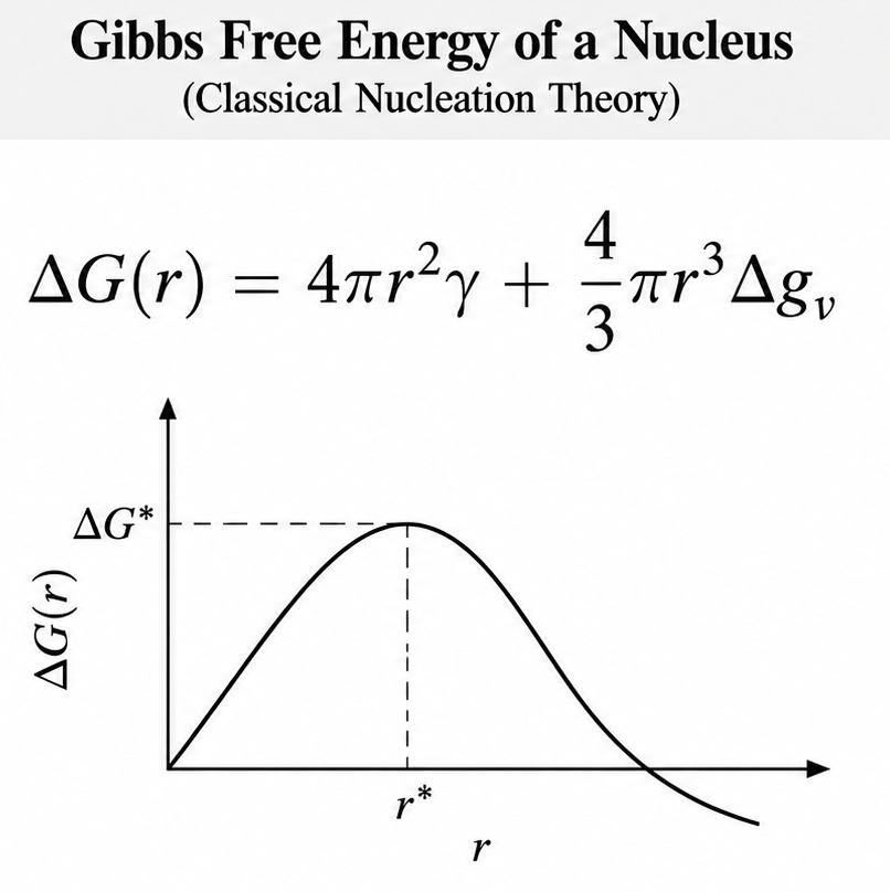
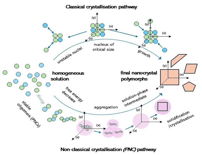
Modern theoretical efforts explaining the origin and development of shape anisotropy under different ligand environments include stages such as differentiated nucleation, cluster aggregation and growth. For ligand-regulated cases, of the two prevalent nucleation theories — classical and non-classical nucleation theory (CNT) — the latter is increasingly gaining evidence.
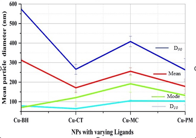
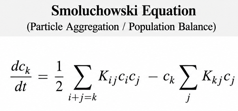
Nevertheless, growth-phase anisotropy is generally explained by oriented attachment and Smoluchowski-type kinetics of cluster aggregation. In the presented case we have an interesting observation: be it α-Fe₂O₃, ZnO or CuO nanocrystals, with methylcellulose as a pure ligand or sugar press mud extract as a complex ligand dominated by the lignocellulosic fraction, the similarity in the shape of the majority populations of the crystals with PDI ≥ 0.7-06 is apparent. This raises a statistical question: why does the non-homogeneous ligand environment not shift the population and the anisotropy index compared with the pure and homogeneous environment? We are seeking this answer from the biomimetic chemistry of mineral synthesis.
Source: L. M. Verma et al., J. Mater. Chem. A 13, 33365–33384 (2025). doi:10.1039/D5TA02013A ↗
Theme C
Crystal Structural Information Transfer
Shape anisotropy
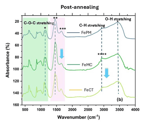
In the bottom-up approach to nanosynthesis — particularly sol-gel, hydrothermal or precipitation methods — ligands are used to define the nucleation and the configurational states of the resulting nanomaterials. But whether the structural/configurational information of the nucleation states is transferred linearly to the final states through successive stages, without loss, is not yet well understood. Studies suggest it may depend on the nature of the ligands, the nuclei and the heating temperature (sintering) used during processing.
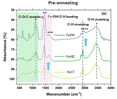
In the particular case of α-Fe₂O₃ under ligands such as CTAB, methyl cellulose (MC) and press mud extract at 500 °C, the structural information on shape remains preserved post-sintering, while the material goes through a phase transition from hydrated forms of iron oxide such as FeOOH to the thermodynamically most stable form, hematite. This preservation of information in the pre- and post-sintering stages hints at the same energy pathways being taken by the system during nucleation and growth in solution, and during Ostwald ripening and coalescence in the sintering phase in the presence of ligands, irrespective of the possible energy landscape of the system. However, it remains unexplained whether this effect arises from the local environment under thermodynamic control or is a result of Wulff construction and an equilibrium shape.
Source: L. M. Verma et al., J. Mater. Chem. A 13, 33365–33384 (2025). doi:10.1039/D5TA02013A ↗
Theme D
Charge Transfer at the Mineral-Water Interface
Light-based processes
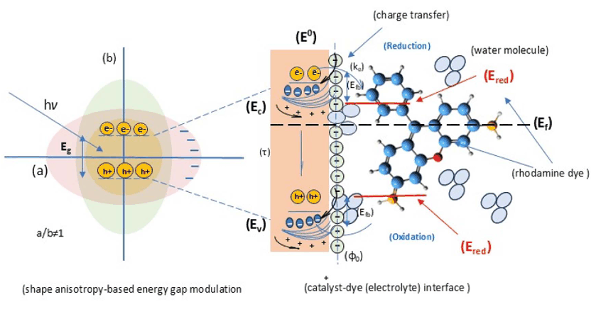
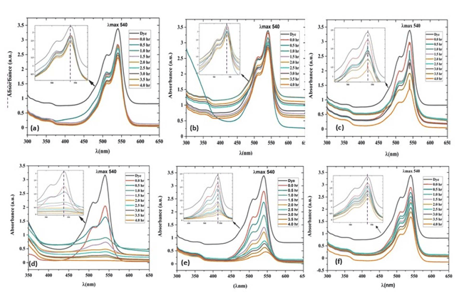
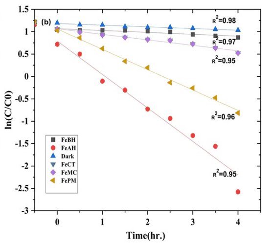
Charge (e⁻) transfer events are at the centre of life and natural processes. Photochemical, electrochemical and biological redox processes are popularly seen as different, but our experiments show they share an essential similarity. For instance, heterogeneous photocatalytic dye degradation involving charge transfer at solid–liquid (semiconductor–dye / mineral–water) interfaces is not operationally different from electrode–electrolyte or enzyme–receptor interfaces.
We have observed in our experiments that light (hν)-induced charge transfer at α-Fe₂O₃, ZnO and CuO/dye interfaces appears to obey the Butler–Volmer equation macroscopically, while obeying the nonadiabatic electron-transfer model (Marcus theory), which was primarily developed for homogeneous molecular electron transfers.
Source: L. M. Verma et al., J. Mater. Chem. A 13, 33365–33384 (2025). doi:10.1039/D5TA02013A ↗
Theme E
Interfacial Charge Transfer Kinetics
Electrochemical descriptors at light-based dynamic interfaces
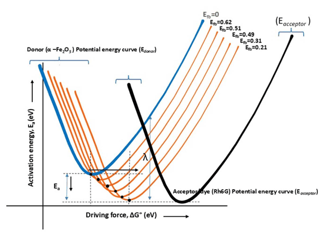
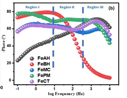
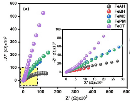
In our particular case of localised semiconductor surface states and a molecular acceptor, the electron transfer rate constants (K_ET) were found to correlate with electrochemical descriptors such as electrochemically active surface area (ECSA), flat-band potentials (Mott–Schottky plots) and charge-transfer impedance (Nyquist and Bode plots) — essentially bridging Butler–Volmer theory (for the electrode interface) with the Marcus framework (for molecular/biological systems), with the evidence found in photocatalysis. However, our experiments have shown that the essentially multistep photocatalytic rate constant (K_p) for dye degradation is fairly explainable by a modified Marcus framework. Research in this direction needs more empirical data for the unification of prevalent theories.
As discussed above, electron transfer at the metal oxide (semiconductor)–dye interface can be explained using the impedance of heterogeneous charge-transfer interfaces (Nyquist and Bode plots) — descriptors traditionally known for the electrode–electrolyte interface. Charge transfer through light (hν)-excited states is increasingly being studied using electrochemical toolboxes. However, we are far from a coherent theory.
Source: L. M. Verma et al., J. Mater. Chem. A 13, 33365–33384 (2025). doi:10.1039/D5TA02013A ↗
Theme F
Nano-bio Interfaces
Nano-biotechnology
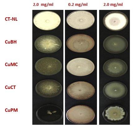
Current research on the nano-bio interface has several dimensions, one of which is how nanoscale materials affect microbial life, such as bacteria and fungi. This is important for the development of disinfectants and new antibiotics, and for the safe use of synthetic nanomaterials. Evidence is growing for distinct effects of materials on bacteria and fungi owing to their nano size and surface charges. However, the mechanism through which these effects operate remains poorly understood.
For instance, our experiments have shown that the same set of nanoparticles (α-Fe₂O₃, ZnO, CuO) capable of initiating redox reactions shows different activity towards different species of bacteria and fungi. This raises interesting questions about the nano-surface–microbial cell wall interface.
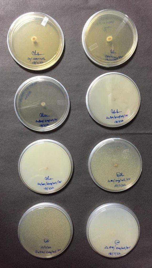
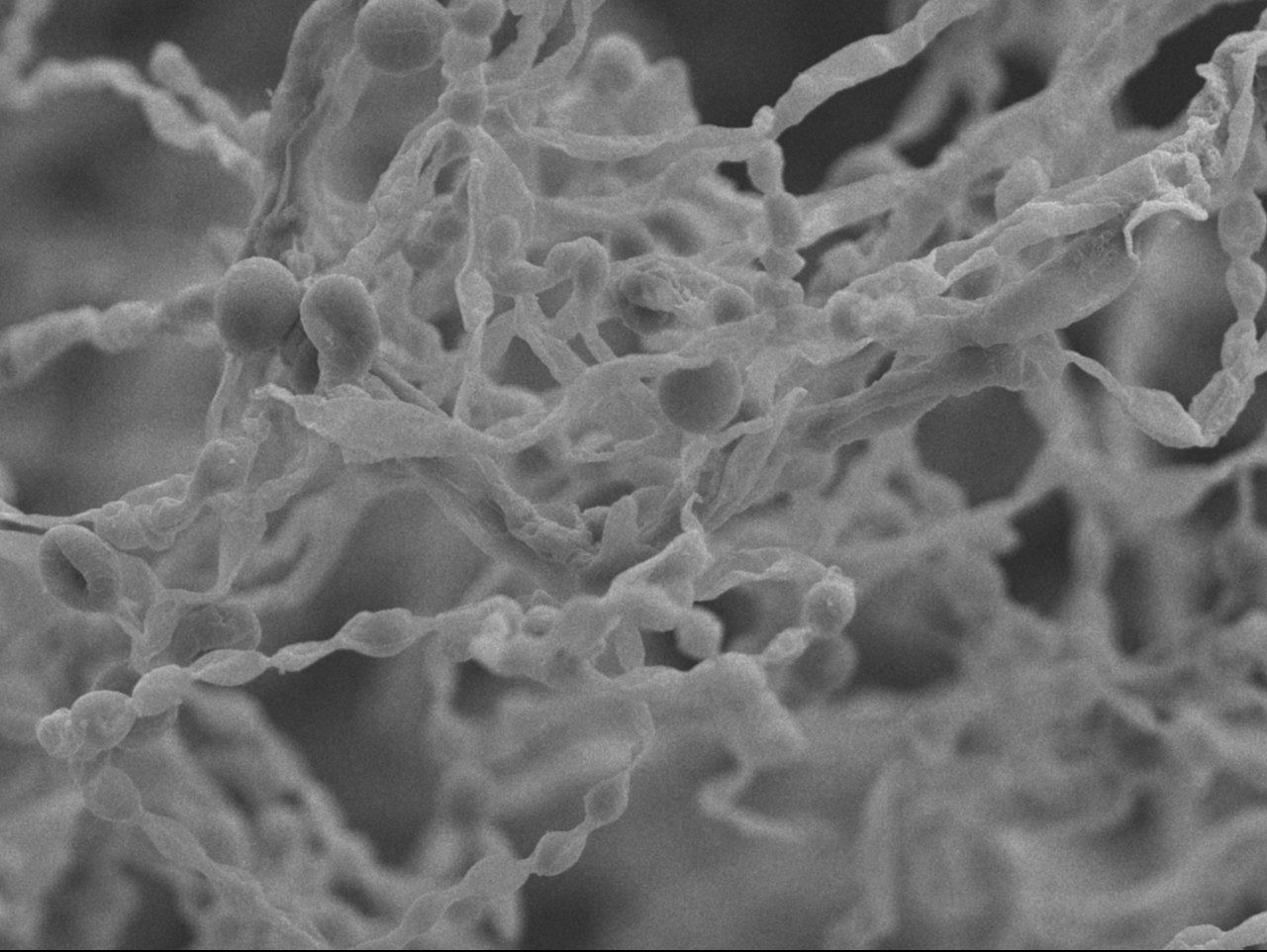
Source: Doctoral thesis work (L. M. Verma).
Theme G
Nutrient Use Efficiency
Soil–plant continuum
We are constantly searching for new materials, particularly from bio-resources/wastes, that can deliver macro-nutrients (particularly NPK) efficiently while avoiding environmental losses. We use bioengineering and biotechnological principles to address the problem of nutrient use efficiency (NUE).
Our primary question in this theme is to find standard materials with an optimum release rate (k_R) and soil diffusion coefficient (D) that can deliver agricultural cargos. We also study soil organic carbon (SOC) in terms of nutrient use efficiency, soil health and climate-resilient agriculture.
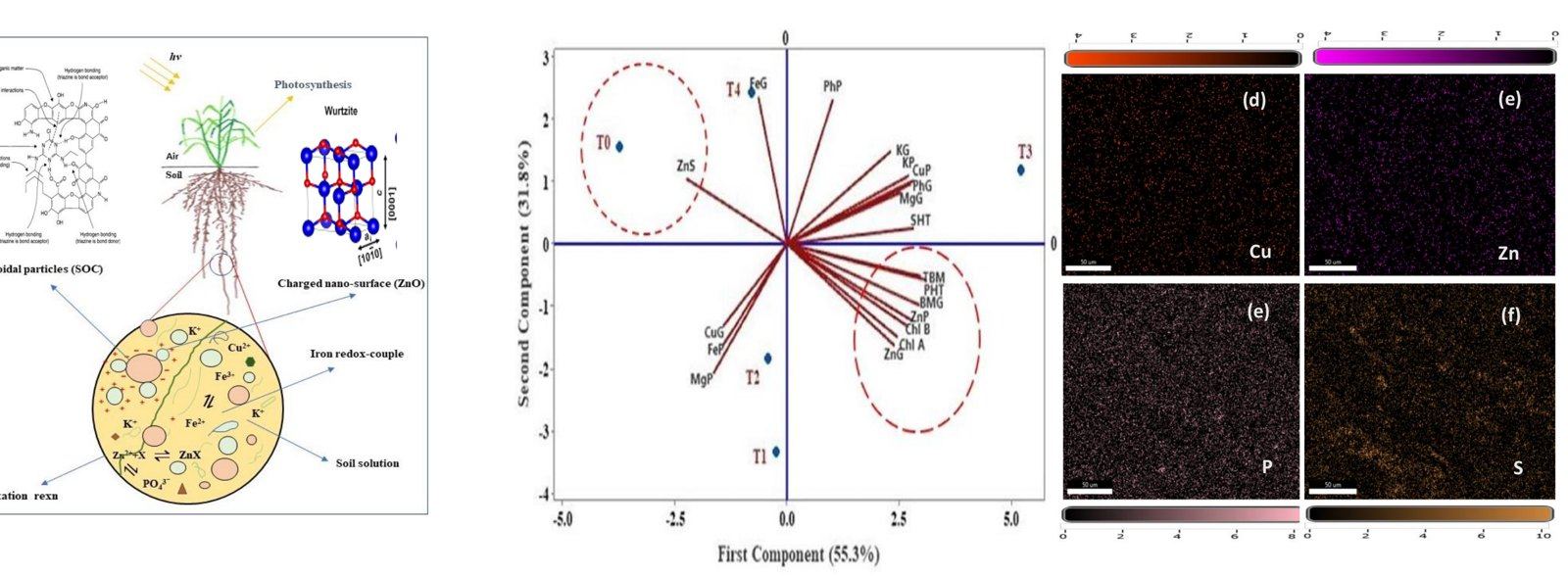
Source: L. M. Verma et al., Sci. Rep. 14, 4074 (2024). doi:10.1038/s41598-024-53682-0 ↗
Theme H
Computational Chemistry & Soil Science
Multiscale modelling and AI/ML
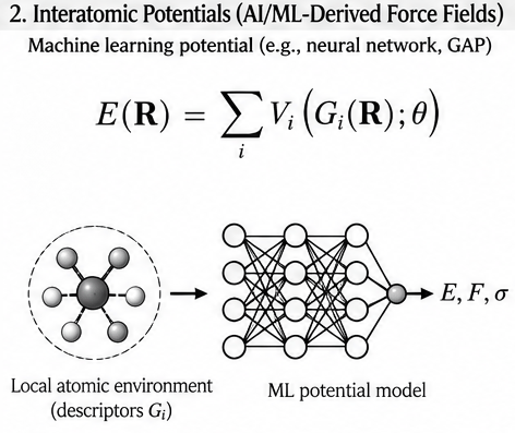
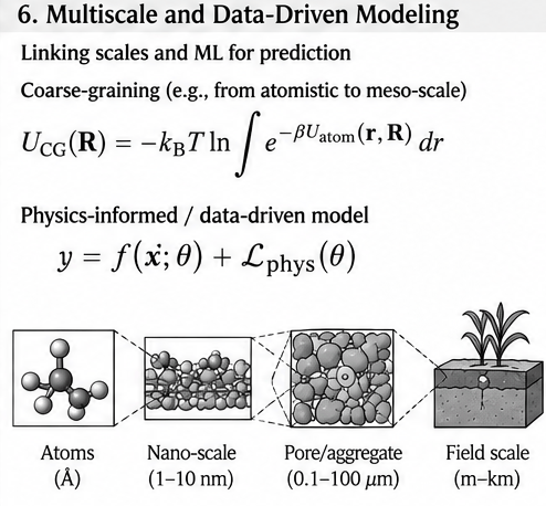
In collaboration with expert groups, we are developing capabilities in multiscale modelling using AI/ML-based force fields to solve problems in materials design, adsorption, release and diffusion in soil matrices.
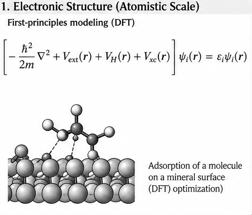
Source: Research overview; in collaboration with the Computational Chemistry Group, IIT Delhi.
Selected publications and full research profile